So hearts come color coded now? Oh man, surgeons have it easy
source: http://www.pediatricheartspecialists.com
I’ve given up on learning the secrets to winning a woman’s heart and settled for learning
how they tick…or should I say beat. Well, here is what I’ve learned so far…
A Word or Two about Gravity
Yes, gravity is always an issue. It acts on blood moving through the body no differently than
it acts on feathers, wooden balls, Chuck E. Cheese tokens or whatever else that 16th century wild man
Galileo decided to drop from tall buildings in Pisa. The heart performs remarkably well
despite blood’s downstream tendencies, pumping against the stream so that no organ gets
cut out of the action.
Hey don’t fall asleep on me yet. I’m only telling you this painfully obvious fact to point out
that since the heart muscle directs blood flow there really is no reason to go looking uphill to figure out where it all begins. Which direction you choose to describe blood flow starting from is really a matter of perspective, by that I mean the fate of oxygenated versus deoxygenated blood. Since it’s all about me, I’m going to start with the flow of oxygenated blood traveling from the lungs.
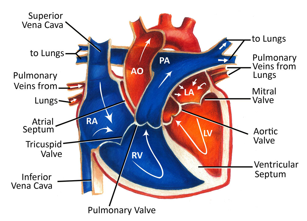
Listed in order
Oxygenated Blood
- Lungs
- pulmonary veins
- left atrium
- bicuspid valve/atrioventricular valve
- left ventricle
- aorta – its branching arteries which feed the systemic circulation
Deoxygenated Blood
- superior & inferior vena cava
- right atrium
- tricuspid valve/right atrioventricular valve
- right ventricle
- pulmonary semilunar valve
- pulmonary artery/trunk
- lungs
The tune goes like this…

I was always too busy pumping the brakes and swearing to notice the striking similarities
So the lungs have done their part, allowing red blood cells to exchange their CO2 from the
tissues for oxygen from the aveoli into neighboring capillary beds. Once oxygenated, the
blood returns to the heart from the lungs by a set of pulmonary veins which empty into the
Left atrium like jumper cables to a battery. Medieval architects constructed atriums in
cathedrals to serve as the first giant, open chambers that you would enter. The atriums of
the heart are constructed in much the same way (that’s right, I learned that in public
school). Blood from the atrium enters into the left ventricle after passing through the
bicuspid valve. As the ventricle contracts the oxygen charged blood moves into the aorta
from which it can flow freely into the wild (or the systemic circulation making up the rest of
the body if you want to be boring). The aorta is a major intersection of arteries that branches
off to the body, the Jersey Turnpike of the vascular system.
So now that the blood has had time to mingle with the organs and tissues, toured the
sights, been places…what happens in Vegas stays in Vegas that sort of thing, it is just
about tapped out of oxygen. The blood then returns to the heart from two different
directions. Blood from the upper body will be entering through the superior vena cava and
the blood coming from downtown will be entering through the inferior vena cava. Much like it did on the left, blood will first enter the right atrium. Blood will then flow into the right
ventricle after it has passed through the double doors of the tricuspid valve. Contraction of
the ventricle will move the deoxygenated blood into the pulmonary artery where it will revisit
the lungs for more of that sweet sweet oxgen. I love a happy ending.
A few things to consider..
Valves are a pretty nifty thing to have in a fluid environment under relatively high pressure. The heart has a lot of blood to move and can’t just take a deep breath and let it all out at once
like the big bad wolf. It needs to take a lot of smaller breaths, relax and contract. The valves
stop the blood we started with from being forced backwards under the pressure every time
the heart muscle changes it’s shape.
The left and right sides of the heart are cleverly separated from each other by septa. These are thick, tough walls of cardiac muscle that keep the oxygenated blood from mixing it up with the deoxygenated blood among the atria and ventricles.
The pulmonary veins are kind of special. If you’ve ever studied the difference between arteries and veins, the veins are typically illustrated in textbooks as blue and the arteries are always red. Illustrators do this to emphasize the oxygenated versus deoxygenated nature of the vessels. So then non-conformists like the pulmonary veins come along and throw everything off because they deliver oxygenated blood to the heart. So now what color should we use?
Anyway, I hope this helps you on your way towards academic rock stardom. Stay classy my friends and never stop learning 🙂
https://forgottenphysiology.wordpress.com/2013/02/18/just-breathe/

Thanks for this blog. I enjoyed your take on blood flow 🙂
Interesting stuff! Your enthusiasm for the topic is clearly on display.