Me Talk Pretty – Nociceptive flexion reflex (NFR)

SU11_GTY_sb10064912a-001_a_hzKnown on the streets as the withdrawal reflex, NFR refers to our body’s involuntary, ninja-quick neuromuscular response to pain. It’s our super sexy physiology term of the week and quite possibly our spiciest one yet. I even like saying it out loud (sometimes in a French accent).

Isn’t it nice to know that our body has our best interests in mind despite our worst judgment? If you’ve ever not been paying attention for a moment and let your hand stray a little too close to a hot stove (like who hasn’t) then it’s highly likely that you’ve witnessed your NFR in action. You probably yanked your hand away at what felt like the same instant you experienced pain. We perceive pain and withdraw from it simultaneously and our body does this seamlessly. If you’re the least bit skeptical just think back to that scolding hot bowl of chili con queso you dropped. YOU didn’t drop it. Your hands let go of it.

What experience could be more direct than pain? In reality, pain may not be as direct a phenomenon as you might think. When it comes to our involuntary withdrawal from pain there are at least 3 stages that occur. Once any part of your body is injured nerves in the vicinity transform that moment into a lightning quick electro-chemical signal. That signal has to take a journey from the site of the injury to the spinal chord. A new signal is sent from the spinal column to the appropriate muscles to flex and manipulate that stray body part away from the pain stimulus. Perhaps one of the reasons this process is so blindingly fast is the fact that the nerve control center in this case is the spinal chord and not the brain.

Let me set the scene…

It’s a perfect July afternoon. The grills are out. The air is saturated in honeysuckle flowers and spicy, charcoal kissed meat. You’re barefoot in the grass, loving life in your favorite shorts and a barbeque stained t-shirt. Oh yeah, you know what I’m talking about. You have a half-eaten bratwurst in one hand. Your other arm is free, innocently reaching out to grab the Frisbee that your friend just tossed to you (you crazy kids). She tossed it high. You take a step back unaware that laying in wait in the grass is a jagged piece of gravel just begging to be stepped on by bare feet. You plant your foot on the rock…

1) Tiny, branching pain receptors suspended within the epidermal layer of your heel, like tree roots, transmit an electrical signal up a long network of neuron fibers that form an intricate junction at the spinal column not unlike a downtown D.C. intersection.

2) Once the message reaches the spinal column an alpha motor neuron sends a signal back down your leg.

3) The signal from the alpha motor neuron reaches a neuromuscular junction in the hamstrings of your leg, initiating their contraction. This flexes the knee, pulling your foot off of the ground to minimize damage to the soft tissue of your foot so you can frolic in the grass another day.

Stay curious, stay classy, and never stop learning my friends 🙂

Fun Facts – A Touch of Gray

ImageOh yes, it WILL happen to you. Don’t worry it happens to all of us, aging that is. More specifically, gray hairs happen. That’s right our bodies, unlike diamonds, aren’t forever. Hair follicles lose their pigment as we age. There is a family of pigments in our bodies called melanins that contribute to hair and skin coloration. So whether your complexion is Godiva, chocolate brown like Djimon Hounsou, salted caramel like Rosario Dawson (call me), or slow churned vanilla like Olivia Wilde, you have melanin to thank. Oh man, all this talk about race makes me want ice cream.

Anyway…

Hair is like sea coral. Yes, that’s random…try to focus. Much of the underlying structure of a coral is the deposited, calcium carbonate remains of dead cells that harden and accumulate while new cells grow over top of the layers. The structural anatomy of a hair follicle is actually curiously complex but the portion of that follicle that we see protruding from the skin (the hair shaft) is just the deposited remains of dead keratinocytes (keratin containing cells). However, unlike coral, the living cells are underneath. Keratin is the crazy strong protein that gives hair, nails, and skin their structure and durability. That same protein that allows Scarlett Johansson’s golden locks to flutter in the wind are what make a rhino’s horns hard enough to dent the door of a Jeep when the tourists get too close.

Clusters of rapidly dividing keratinocytes at the root of the follicle team up to produce keratin. Cell division and keratin production creates a dense mass of material that builds from the bottom up. Meanwhile, bordering those feisty root cells are melanocytes that pass their melanin granules to the keratin producing cells. So even as the cells die they retain whatever color was passed on.

As we age melanocytes become less active and die off and so more and more hair shafts reach the surface without pigment. So just to be clear, your body doesn’t produce gray hairs. It simply stops producing melanin pigment within the hair follicles. The resulting hair strand is actually colorless.

Now the rate at which our hair loses its color is largely genetic. In know, genetics seem to be the new cop-out answer. It’s like when they use “instinct” to explain why ducks fly south for the winter. However, this time we have to point the finger at inheritance for our sexy, silver sheen.

Stay classy, stay curious, and never stop learning my friends :-)

The Meninges and the Deadly “Itis”

Your Meninges and You

"Whoah, play that back..whose heart is gonna stop?"

“Whoa, play that back…whose heart is gonna stop?”

It doesn’t seem fair about the brain does it…all the E-Hollywood, red carpet attention it gets? I mean there isn’t a single one of us out there in this crazy world that can survive if our heart stops beating and that’s just as true for snow leopards, crickets, Glen Beck, and 3-toed sloths as it is for foxy human-beings like you and me. However, the brain is treated like the commander and chief of the body (which it pretty much is). By that I mean consider the level of security and protection the body invests in its head of state. Not only is some of the densest sections of bone in the body (contained in the skull) there to protect it from impact like a souped up, Seattle Seahawks NFL helmet but there is layer upon layer of security measures in place to maintain this safe, warm, cozy, zen-like environment surrounding the brain and spinal chord as well. One of those layers of security is a super fancy barrier of connective tissue between the brain and skull referred to as the “meninges.”

Your meninges has 3 layers of its own, the dura mater, arachnoid mater, and subarachnoid space, which all kind of sound to me like strange galaxies of the Star Trek universe in old episodes of the Next Generation. (just saying, I’m a nerd like that)

Oh yeah, that's what I'm talking about.

Oh yeah, that’s what I’m talking about.

No, I’m not going to compare them to layers of a cake. We all know that’s really gross, but it is a fairly dynamic, intricate space. The Dura mater is the outermost layer. It’s thick, tough stuff just like you would imagine the outer layer of a barrier to be. Just beneath the dura is a single layer called the arachnoid mater that has thousands of spindly little projections that connect to the layer below, like scaffolding to a building under construction. The third layer is really more of a space, the subarachnoid space, that contains blood vessels, arachnoid spindles, and cerebrospinal fluid that basically coats the brain and spinal chord.

MeningesBlum

the subarachnoid space is a mesh of branching vasculature within a matrix of CSF fluid

Meningitis

Just like other tissues of the body the meninges is susceptible to infection and can become infiltrated by pathogens like bacteria, viral particles, or any other bad boy microbe within our circulation that makes it past our immune system or the blood brain barrier. When this happens the meninges becomes inflamed (e.g. Meningitis) and it’s about as bad news as bad news gets. Picture the meninges as a continuous inflatable mattress wrapped around the brain and spinal cord that suddenly gets blown up, squeezing and pressing on the delicate wiring of your central nervous system. Not exactly the best summer ever.

Hey, don't get distracted. I'm talking about meningitis here

Hey, don’t get distracted. I’m talking about meningitis here

Meningitis sufferers typically complain of a biggie-sized headache coupled with neck stiffness along with a fever. Other symptoms mimic that of a migraine…light sensitivity, confusion, disorientation, and a general sense of “something just ain’t right!” Now meningitis is an inflammatory process and you know that there are about a bazillion microbes out there that can trigger an inflammatory response upon infection. So when a patient, or even worse, when multiple patients display clinical features of meningitis the clock is ticking for medical detectives to seek out the “infectious agent.”

The Usual Suspects

N. meningitidis, pain in the butt, gram negative staining diplococci that looks like tiny coffee beans under the scope

N. meningitidis, pain in the butt, gram negative staining diplococci that looks like tiny coffee beans under the scope

When we want to point the finger at bacterial causes of Meningitis three misfits come to my mind almost immediately; Neisseria meningitidis aka meningococcus, Streptococcus pneumoniae aka pneumococcus, and Hemophilus influenzae. All three of these shady characters like to hang out in the nasopharynx of our upper respiratory tract. So they typically spread from infected individuals by means of “respiratory secretions” as in kissing, coughing, and sneezing. Of course, if the infected is an infant the mode of transmission can include just about anything they can put in their mouths and pass on. S. pneumoniae and H. influenzae are opportunistic little bastards. That means don’t go spraying everything and everyone down with lysol just yet, because these guys typically only spring into action when we are already immunocompromised in some way. Meningococcus, on the other hand, is the only known bacteria to cause meningitis on an epidemic level.

You may have noticed that I didn’t dive into viral causes of meningitis. Are you kidding..that list is like 3 pages long! Viruses pose an even greater threat in that they are much much smaller, mutate at the drop of a hat, and have more modes of transmission than…Lil Jon has bling.

Lil+Jon

Exactly, more than that.

A lumbar puncture is typically the diagnostic procedure of choice when meningitis is suspected. This undeniably scary, long needle is inserted into the patient’s back to extract Cerebrospinal fluid from the spinal canal, usually through access to the lumbar vertebrae. Remember that your brain and spinal chord are suspended in fluid 24/7 that should be vacant of things like, viruses, bacteria, and even your own blood cells. So those few precious drops of CSF are like diagnostic gold to the folks working the clinical lab. They will make microscope slides to look for white cells and microbe hitchhikers and perhaps most importantly culture that CSF sample on clean, nutrient rich agar plates to see what pathogens grow. Many times they can positively ID the culprit like a closed case file in an episode of “The First 48.”

Stay curious, stay classy, and never stop learning my friends 🙂

Me Talk Pretty-blood brain barrier

Hello! No, you look great today.

This week’s super sexy, fancy pants physiology term is…

Blood brain barrier

Every organ in your body is supplied in some fashion with blood. Yes, I know I haven’t said anything too shocking yet. However, as far as
the body is concerned the brain is like Queen Mother and requires the extracellular fluid (fluid without the cells) to be separated from the circulating whole blood by a royal guard of specialized endothelial cells. Just like I pointed out in another article, True Blood, blood is a pretty wild mixture and not everything in that mixture is useful or even safe for the precious, fine tuned neurons of the Central Nervous System (CNS). A network of painfully tiny capillaries form an intricate spider web mesh that filters out big nasty things (microscopically speaking) like bacterial cells, microscopic fungi, and cellular debris as well as large hydrophyllic molecules like proteins, and a buffet of potentially bad news chemical compounds.
The Blood Brain Barrier has the mad skills to allow vital compounds like CO2 and O2 to slip through while actively transporting (uses membrane protein channels) glucose, hormones, and amino acids. Remember that the blood vessels of the body have elastic qualities and those vessel walls become more permeable (leaky) during inflammation.
When doctor’s perform spinal taps, drawing up CSF (cerebrospinal fluid) from within the patients spinal column with a syringe, the
fluid tells a story. In healthy patients spinal fluid should be a ghost town…clear, colorless, and devoid of red cells, white cells, and just about everything else. Patients with active infections of their CNS may show white cells, red cells, increased protein, glucose (like in Tuberculosis cases) and discoloration of the fluid sample. The inflammation or infection in that case has overwhelmed the BBB’s ability to filter. Sometimes you truly understand the value of a thing when it stops working.

Stay curious, stay classy, and never stop learning my friends 🙂


That Dizziness Thing

spinning-merry-go-round_medium

After two pink bags of cotton candy all the rides were a little blurry that day.

Ok brace yourself, because I’m about to make a painfully obvious statement. Sometimes what we say we’re feeling and what we are actually feeling is not the same thing. Yes, you do it too. Oh yes you do! Do I need to call your mom and let her know your pants are on fire? Listen, I don’t want to fight about this, but I will say that there are a handful of terms good people like you and I use to tell our doctors what we’re feeling because we don’t know any better. For example, when was the last time you felt dizzy? Perhaps I should say lightheaded, or that you felt like you were going to faint (presyncope). Maybe you felt like the room was spinning (vertigo).

We use “Dizzy” to describe all of these sensations when in fact sometimes we’re only experiencing one of them. When the teenager behind the counter at the ice cream shop takes your order you don’t say “give me two scoops of the really cold kind” do you? Sometimes names matter. Well get ready, because we’re about to get technical.

big-az-balloons-3

Hey Buddy, maybe you should sit down. You look like you need a juice box.

Light headedness is typically the result of a sudden drop in blood pressure (hypotension). Our bodies are a lot like one of those giant, dancing balloon characters outside of a used auto dealership. You cut off the fan (the air flow) and the balloon man falls to the ground. Well when our fan shuts off (our blood flow to the head is restricted) we faint. We refer to this as “syncope.” That feeling that we are about to faint is “presyncope.” We can also experience the lightheaded blues when we stand up too fast and we refer to this as “orthostatic hypotension,” where the blood flow is affected by the positioning of the body. Ok blah blah…that’s all very interesting (yawn), but I want to get to the good part, vertigo.

Vertigo is CRAZY interesting. Actually, for the rest of this article I’m just going to talk about vertigo. Everything else is dead to me.

Vertigo is a profound sensation of disorientation. You can have the feeling that the room is spinning or that you are spinning. You can also experience the sensation of falling or that the ground or room is tilting. All of these trippy sensations can make you feel incredibly off balance and often lead to nausea.

All of these sensations are caused by a malfunction (minor or severe) in the way your brain is receiving information from the balance organs of the body. What kind of malfunctions you ask? Well if the possible causes of vertigo were a dusty road in New Mexico that split in two, one road sign would read “sensory neural pathway” and the other would read “organs of the inner ear.” Need more? As the comedian Bernie Mac used to say, “Let me break it down for you like a fraction.”

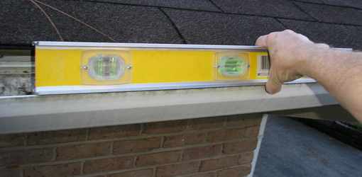
Your brain talks to your body constantly. Like an overprotective mother the brain asks your eyes “where are you? What are you doing right now? What do you see?” However, Mother is just looking out for us, taking that visual input and coordinating our skeletal muscles/motor control to maintain an upright posture and orient the fluid within our inner ear like the bubble in a carpenter’s level (see fig. 2) so that we have an accurate perception of up and down, left and right, backwards and forwards. That intimate communication between brain, eyes, and motor control is referred to as the vestibulo-ocular reflex. Go ahead and click this link

https://forgottenphysiology.com/2013/12/19/me-talk-pretty-vestibulo-ocular-reflex/

it won’t bite 🙂

Balance is in your ears?

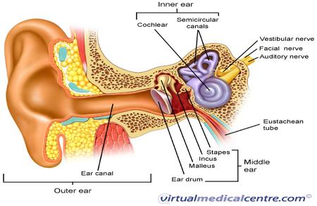
This balancing act is brought to you by our inner ear. The physical interaction of sound waves within the intricate, spiraled canal of bone called the cochlea in conjunction with the stimulation of auditory nerves are largely responsible for how we hear sound. However, one of the more mystifying tricks our inner ear can perform is that of our perception of balance. Alright, we need to zoom in on this thing. Look down there…

2191_ear_anatomy_450

ok keep your eyes on the swirlie, looping purple thing (labyrinth of the inner ear) in the diagram. This is where the magic happens.

No no, I mean closer than that…

inner-ear

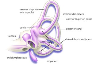
Welcome to the Labyrinth of the inner Ear

how-clean-repair-gutters-5

A special thanks to Thing for demonstrating the Carpenter’s level

This structure has more twists and turns than a back road in Lexington, Virginia so I’m only focusing on two main parts for now, the semicircular canal and the otoliths. That reminds me, how familiar are you with building houses? That’s ok, just look to the right of the screen. See that’s a carpenter’s level. Builders set it on top of fence posts, center blocks, steel support beams and anything else they need to have level with the ground. That bubble in the center tells them how far off they are. Inside each ear are three very similar structures, semicircular canals, made from bone and tissue that your brain communicates with to maintain rotational balance and orientation (like when you’re busy doing back flips like Night Crawler from X-men). These are tiny, fluid filled canals that loop parallel and perpendicular to each other. That itty bitty amount of fluid inside pushes on the hairs of a structure called a cupula that translates that mechanical movement of the fluid into an electrical message that tells the brain something like “hey, this guy’s doing a cartwheel.” You have three of these in each ear; anterior, posterior, and horizontal that correspond to vertical, horizontal, and diagonal rotations of the body.

The more you learn about the structures of the inner ear the more this all begins to sound like some freaky game of pinball, but we’re not done. The otolithic organs allow us to sense linear acceleration by aid of tiny crystals suspended in viscous fluid [I always thought the term viscous was kind of creepy, but that’s just me]. We have two otolithic organs on each side; the urticle, and the saccule. The urticle is tuned-in to horizontal movement and the saccule is all about the verticle changes in position.

The otolith organ is this crazy looking sac of fluid with a cluster of sensitive hairs. The fluid as I mentioned before is thick, viscous and contains a lot of tiny calcium carbonate crystals that make it grainy as well. So when the head moves this nasty substance produces friction against the hairs and that inertia is translated into vertical and horizontal linear movement by the brain.

Back to Vertigo

When you are suffering from vertigo disruption of the balance organs (especially those of the inner ear) and the sensory neural pathways that talk to those organs are typically suspected. Benign paroxysmal positional vertigo (BPPV) is common when there is inflammation from infections or injections. If inflammation is significant enough it can disrupt the movement of otolith crystals within the inner ear, sending mixed signals to the brain. That can definitely make you feel like you went too many seconds on the mechanical bull in Salt Lake City.

Brother, what a night it really was...

Brother, what a night it really was…

Stay curious, stay classy, and never stop learning my friends 🙂

Me talk pretty – Vestibulo-ocular reflex

Hello and welcome to the first segment of “me talk pretty” where I bring you tongue twisting terms from the most remote corners of physiology. This week’s super sexy, fancy-pants physio term is….

“Vestibulo-ocular reflex (VOR)”

This is a highly adapted eye reflex in humans that keeps images fixed or stablized on the retina (in the center of your visual field) by allowing the eyes to move in the opposite direction of head movement like a compass needle that points to magnetic north no matter how much the ship tosses on the waves. Your VOR is incredibly fast (fraction of a second) and it needs to be in order to compensate for even the most subtle head rotation along an axial plane (vertical, horizontal, or diagonal). It is what makes clear vision possible. If you were a catcher for the Yankees and your VOR were a fraction of a second off you would be grabbing for blurry fast balls at 90+ mph, but hey, who wants to live forever? (No one should be grabbing for blurry balls) So the next time you’re trying to read your Vanity Fair magazine on a Chinatown bus with bad suspension, finish a sudoku puzzle on a plane while someone’s devil spawned child is throwing a fit and kicking the back of the seat, or just trying to check out the fit brunette jogging by out of the corner of your eye, remember to thank your VOR for keeping that image nice and clear.

Hiccups – the Devil’s plaything

devill

Is that the same devil you had in mind? Twins!!

There’s public radio telethon week, toll roads, vending machines that give you Diet Pepsi when you clearly pushed the “Coke” button, and near the top of the list are hiccups. These are a few of my least favorite things. Oh, you know this guy loves the human body. There are few marvels in this world more elegant, more profoundly complex than the body and it’s nuts and bolts, but son of a [CENSORED], hiccups are the devil! I seriously doubt that a successful pick up line has ever been delivered while battling with the hiccups. Hiccups lie in wait like mean-spirited hecklers waiting to kill the mood of your acceptance speech or interrupt the punch line of your best dirty joke. So in this episode of Forgotten physiology we’ll pose the question; Hiccups, what’s your deal?

Yes, I prefer to think of my hiccups as the old men from the muppet show
http://www.youtube.com/watch?v=PGfx3QAV64M

Synchronous Diaphragmatic Flutter (or Miss Jackson if you’re nasty)

Mechanically speaking, hiccups are nothing more than spasms of the diaphragm; short, involuntary contractions forcing air through the windpipe that is then immediately closed off by the tough, elastic flap of the epiglottis. This closes off the vocal chords like a trap door that says “No more air for you! No!” and produces that signature, sexy frog sound of a classic hiccup.

KF5015

The phrenic nerves begin in the cervical vertebrate (C3-C5) and zip line down between the heart and lungs to connect with the diaphragm.

You have a left and right phrenic nerve that connect to the diaphragm to control its contractions and receive sensory input in return. “Sensory input?” Well sure, you don’t just blindly send out messages. You look for a response. Your nervous system is all touchy feely and needs to be able to sense where everything is…no organ left behind. Those nerves are the only electrical wiring operating the movement of the diaphragm. So any significant disruption or irritation of the phrenic nerves can cause spasms and ultimately hiccups. For example, if you just had a bad breakup and decided to go Tazmanian Devil on 2 large orders of pork fried rice from Wong’s Wok, a full or distended stomach can press on the phrenic nerves and trigger hiccups.

But who is really in control?

We may not think about it but the diaphragm is made of “skeletal” muscle. If you remember our chat from muscle mania, skeletal muscle is under the control of the somatic nervous system. This typically means that it is under our control. We can, for example, contract our diaphragm muscles ourselves and draw in a deep breath. However, we don’t exactly tell our diaphragms to spasm do we? Ahh…here’s where it gets interesting.

A thought experiment..

You and your body are one and the same right? I mean you don’t feel separate from your body do you? If I ask you to pass the mashed potatoes you don’t ask your hand to grab the bowl. Your brain and your hand flow together, without interruption.
Now think about a hiccup. Does that ever feel like something YOU did? No way, the hiccups happen as reflex and your brain gets the feedback. These are controlled by a “reflex arc,” neural pathways (composed networks of neuron cells) that connect to the spinal chord BEFORE reaching the brain. Some neural pathways have a long way to go to reach the brain, your central command center. The spinal chord works like an intermediary traffic control center, managing the local stuff and all the sensory input from the extremities. This allows motor reflexes to occur instantaneously without checking in at the office (brain) first. Your brain isn’t left out of the loop. The motor neurons of the spinal chord are simply the first to respond.

But why me? Why?!!!

Typically when we get the hiccups we were eating too much or eating too fast, drinking too fast, or hitting the sauce a little heavy (either alcohol or carbonated beverages). Episodes can also be brought on by excitement. I’ve always said that too much happiness is dangerous. These hiccups don’t last long and typically resolve on their own without tongue pulling or drinking water upside down.

There are of course persistent hiccups that are linked to an encyclopedia worth of clinical problems ranging from stress to heart issues to neurologic and metabolic disorders. So if 48 hours have gone by and you’ve still got the chirps then it’s probably a good time to see your doctor my friend.

Stay curious, stay classy, and never stop learning my friends 🙂